Our experienced team of radiologists and technicians provides Ultrasound-Guided (USG) biopsies to assist in diagnosing a wide variety of conditions. Using real-time ultrasound imaging, we precisely guide the biopsy needle to collect a tissue sample from the targeted area, ensuring minimal discomfort and maximum accuracy.
USG-guided biopsies are typically performed to evaluate:
This outpatient procedure usually takes between 30-60 minutes and is performed under local anesthesia, minimizing any pain or discomfort. Most patients can resume normal activities shortly after the procedure. With the help of our cutting-edge imaging technology, we ensure a high level of precision and safety, making the biopsy process as comfortable and efficient as possible.
Our expert radiology team employs advanced CT (computed tomography) imaging to guide biopsies with precision, aiding in the diagnosis of a wide range of medical conditions. By using real-time CT scans, we can accurately identify and obtain tissue samples from challenging or hard-to-reach areas within the body.
CT-guided biopsies are frequently performed to evaluate:
This minimally invasive procedure is typically completed on an outpatient basis and generally lasts between 30 to 60 minutes. Local anesthesia is administered to ensure patient comfort, and most patients can resume their regular activities shortly after the procedure. Our highly trained, board-certified radiologists utilize cutting-edge imaging technology and modern techniques, ensuring both an accurate diagnosis and a safe, comfortable experience. Additionally, CT-guided biopsies provide an enhanced view of complex or deeper tissues, offering a higher success rate in obtaining viable tissue samples.
Fine Needle Aspiration Cytology (FNAC) is a minimally invasive diagnostic procedure that involves collecting cell samples from suspicious lumps, masses, or lesions for further examination. Using advanced imaging techniques, our expert radiologists and cytopathologists ensure that the procedure is performed with precision and accuracy.
FNAC is typically used to investigate:
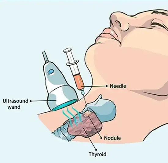
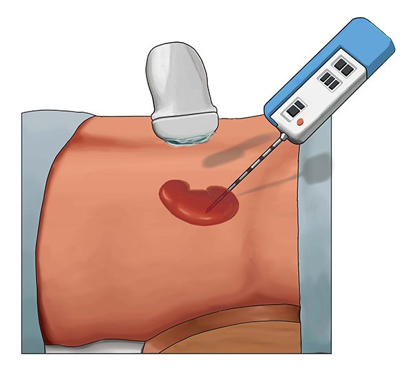
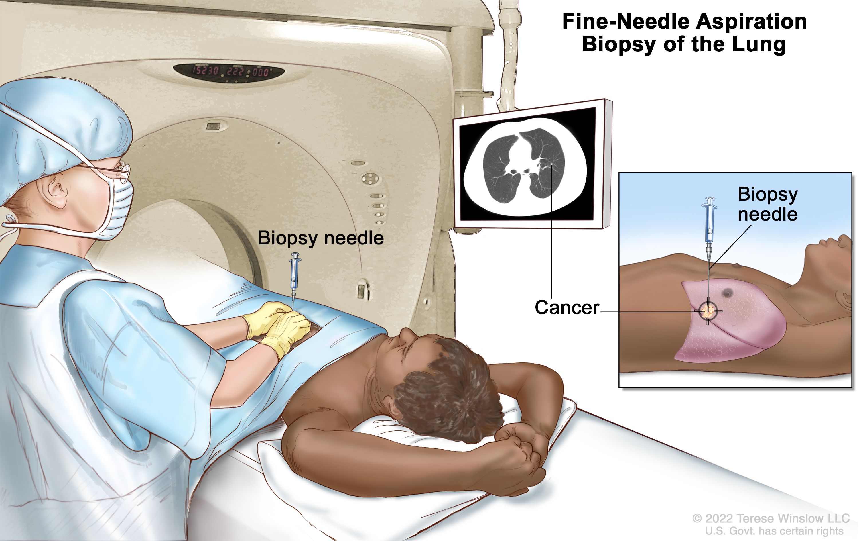
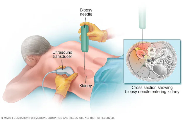
During the FNAC procedure, a thin, hollow needle is inserted into the area of concern, guided by imaging methods like ultrasound or CT scans, to precisely collect cell samples. The samples are then prepared and analyzed under a microscope to identify any signs of malignancy or other conditions. FNAC is a quick, safe, and virtually painless procedure, often performed on an outpatient basis. It typically requires no recovery time, allowing patients to resume their normal activities right away. This technique is highly valuable in providing immediate diagnostic information, which helps healthcare providers determine the most appropriate course of action and treatment plan. FNAC is a cost-effective and reliable alternative to more invasive biopsy methods, offering quick results with minimal discomfort.
Our skilled interventional radiology team specializes in the placement of pigtail catheters to manage fluid buildup or pressure within the body. Using precise imaging techniques, we can insert these flexible, small tubes to effectively drain accumulated fluids or relieve obstructions, promoting recovery and comfort for our patients.
Pigtail catheters are commonly utilized for:
This procedure is typically performed on an outpatient basis, with the patient receiving local anesthesia and mild sedation to ensure comfort. Using imaging guidance, such as ultrasound or CT, the radiologist will make a small incision and guide the pigtail catheter into the desired area. The procedure usually lasts between 30 and 60 minutes, with the catheter remaining in place for the necessary duration, allowing continuous drainage and symptom relief. The catheter may remain in place for several days or weeks, depending on the condition. After the procedure, our team will provide comprehensive instructions for proper catheter care, including how to monitor for potential complications, ensuring a safe and effective recovery process.
Our experienced radiology team uses advanced imaging techniques to perform kidney biopsies, enabling precise tissue sample collection for the diagnosis of various kidney conditions. This minimally invasive procedure helps in making informed decisions for effective treatment.
Kidney biopsies are often used to assess:
The biopsy involves guiding a fine needle into the kidney using ultrasound or CT imaging. This quick, outpatient procedure is performed with local anesthesia, ensuring minimal discomfort. Most patients can resume their daily activities shortly after the procedure.
Our skilled interventional radiology team specializes in the precise, minimally invasive drainage of liver abscesses, using advanced imaging techniques. This procedure helps eliminate infected fluid and accelerates recovery, often eliminating the need for surgery.
Liver abscesses can arise from bacterial, parasitic, or fungal infections and may also result from complications of other medical conditions. Timely drainage is essential to alleviate symptoms and prevent further infection. By utilizing ultrasound or CT imaging, we carefully guide a catheter to the abscess site to drain the fluid effectively.
This procedure is performed on an outpatient basis, with local anesthesia and light sedation to ensure patient comfort. Most patients experience minimal discomfort and can resume normal activities within a few days. The catheter typically stays in place for 1-2 weeks to ensure complete drainage. With years of experience, our expert radiologists use the latest technologies to ensure optimal results and minimal risk of complications.
Our expert interventional radiology team specializes in the placement of permanent hemodialysis catheters, commonly referred to as Permcaths, providing a long-term solution for patients who require consistent dialysis access. This minimally invasive procedure ensures reliable, easy access to the bloodstream for dialysis treatments.
A Permcath is a small, flexible catheter inserted into a large vein, typically in the neck or chest. It offers a convenient and effective alternative to repeated needle insertions, ensuring patients can undergo hemodialysis without discomfort or complications.
Using advanced imaging techniques like ultrasound and fluoroscopy, our radiologists precisely position the Permcath to the most suitable vein for optimal blood flow and accessibility. The procedure typically lasts less than an hour and is performed with local anesthesia to ensure comfort. Once in place, the Permcath can remain functional for several months or even years, minimizing the need for additional procedures.
Our skilled interventional radiology team specializes in the precise placement of central venous catheters, also known as central lines, providing essential vascular access for various medical treatments.
Central lines are crucial for:
Using advanced imaging techniques like ultrasound and fluoroscopy, our radiologists accurately position the catheter in a large vein, typically in the neck, chest, or groin. This minimally invasive procedure typically takes under an hour, with local anesthesia for comfort. Once placed, the central line can remain in position for as long as necessary, ranging from days to months, based on the patient’s treatment plan. Our team will guide you through proper catheter care and provide instructions to minimize potential complications.