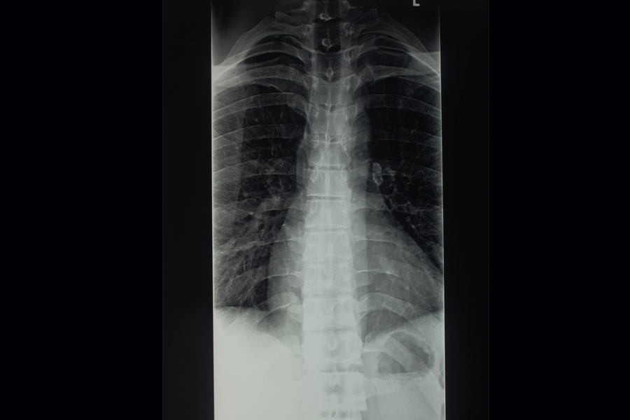
A thoracic spine digital X-ray is a diagnostic imaging procedure used to obtain detailed pictures of the thoracic spine, which is located in the mid-back region, between the cervical spine (neck) and the lumbar spine (lower back). This imaging technique is commonly employed to assess and diagnose a variety of conditions affecting the thoracic spine, including fractures, herniated discs, scoliosis, kyphosis, and other abnormalities. The procedure involves positioning the patient in different angles to capture images from multiple viewpoints, such as front, side, and oblique perspectives. These images enable healthcare professionals to evaluate the alignment of the vertebrae, detect any signs of injury or degeneration, and plan the most appropriate treatment.
Digital X-rays are non-invasive, fast, and provide high-resolution visuals that can be easily shared and interpreted by healthcare providers. This method offers an efficient way to obtain clear images for accurate diagnosis and treatment. Furthermore, the digital format eliminates the need for physical storage, making the results more accessible and convenient for follow-up care and consultation.